 Vikas Diagnostics Best Diagnostics Lab In Kanpur
Digital Barium Studies/ Digital I.V.P.
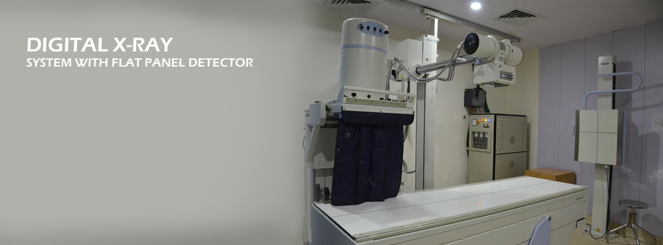
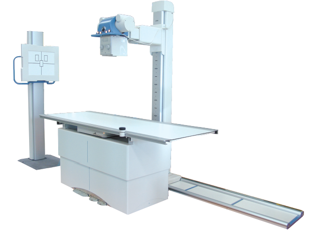
Digital X-ray/Contrast Studies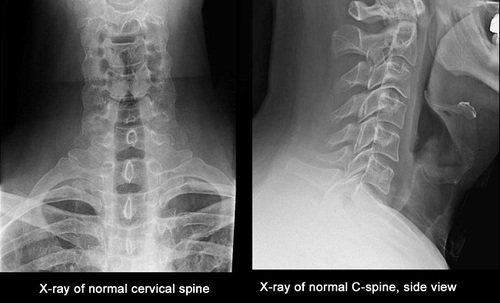
Digital X-ray has been called one of the most significant advances in medical history. It is used in many different ways in medical diagnosis. Routine X-rays involve exposing a body part to a small dose of radiation to produce an image of an internal organ. An X-ray image is produced when a small amount of radiation passes through the body and strikes a sheet of sensitive film placed on the other side of the body. This film is then either placed in a developing machine, to produce images much like negatives from a 35-mm camera, or is digitally stored on a computer.
The most common use of X-rays is to identify and treat bone fractures. It is particularly beneficial in emergency situations. X-ray images of the skull, spine, joints and extremities can show even very fine hairline fractures or chips. After treatment, a bone X-ray can be done to ensure that the fracture has been properly aligned and stabilized for healing. X-rays can also be used to diagnose and monitor the progression of degenerative diseases such as arthritis. They play an important role in the detection and diagnosis of cancer, as well, although CT, MRI and PET are usually better at defining the extent and nature of suspected cancer.
X-ray is a fast and easy procedure. Patients will experience no discomfort or side effects from their examination and are allowed to leave immediately following their X-ray test.
Contrast Studies
Espohagram
An examination of the pharynx (throat) and esophagus using still and fluoroscopic X-ray images. The X-ray pictures are taken after the patient drinks a solution that coats and outlines the walls of the esophagus (also called a barium swallow).
Upper GI Series
A series of X-rays of the esophagus, stomach, and small intestine (upper gastrointestinal, or GI, tract) that are taken after the patient drinks a barium solution. (Barium is a white, chalky substance that outlines the organs on the X-ray.)
Small Bowel or Small Intestine Series
A series of X-rays of the part of the digestive tract that extends from the stomach to the large intestine.
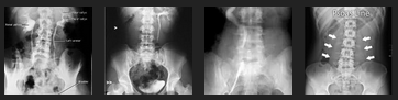
Barium Enema / Lower GI Series
A series of X-rays of the lower intestine (colon) and rectum that are taken after the patient is given an enema with a white, chalky solution that contains barium. The barium outlines the intestines on the X-rays. These X-rays permit the detection of colon and rectal abnormalities including diverticulosis, diverticulitis, abnormal colon movement, dilation (widening) of the colon, polyps and cancers of the colon and rectum. Air can be instilled into the colon along with the barium contrast medium to further define structures of the large bowel and rectum. Polyps and small cancers are more readily found using this method which is called an air contrast barium enema or a double-contrast barium enema. This is the only kind of barium enema that is appropriate for detecting colorectal polyps and potentially curable colorectal cancers.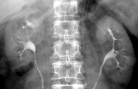
Intravenous Pyelogram (IVP)
An X-ray examination of the kidneys, their drainage to the bladder, and the bladder.
Hysterosalpingogram
X-ray of the uterus and Fallopian tubes; usually done in diagnosing infertility to see if there any blockages.
Arthrogram
X-ray of a joint after the injection of a contrast medium to more clearly visualize the joint.