 Vikas Diagnostics Best Diagnostics Lab In Kanpur
MR Mammography (Breast MRI)
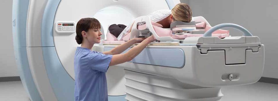
What is MRI of the Breast? 
Magnetic resonance imaging (MRI) is a noninvasive medical test that physicians use to diagnose medical conditions.
MRI uses a powerful magnetic field, radio frequency pulses and a computer to produce detailed pictures of organs, soft tissues, bone and virtually all other internal body structures. MRI does not use ionizing radiation (x-rays).
Detailed MR images allow physicians to evaluate various parts of the body and determine the presence of certain diseases. The images can then be examined on a computer monitor, transmitted electronically, printed or copied to a CD or uploaded to a digital cloud server.
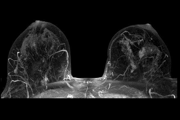
MRI of the breast offers valuable information about many breast conditions that cannot be obtained by other imaging modalities, such as mammography or ultrasound.
What are some common uses of the procedure?
MRI of the breast is not a replacement for mammography or ultrasound imaging but rather a supplemental tool that has many important uses, including:
- Screening in women at high risk for breast cancerFor women at high risk for breast cancer, typically because of a strong family history, MRI may be an appropriate tool to screen for breast cancer. A strong family history is usually a mother or sister who has had breast cancer before age 50. It can also be aunts or cousins, including those on your father’s side. Relatives who have had ovarian cancer also increase your risk. Your radiologist or primary care doctor can look at your family history and determine if screening MRI may be appropriate for you. Depending on your family history, genetic counseling may also be recommended.
- Determining the extent of cancer after a new diagnosis of breast cancerAfter being diagnosed with breast cancer, a breast MRI may be performed to determine:
- how large the cancer is and whether it involves the underlying muscle.
- if there are other cancers in the same breast and whether there is an unsuspected cancer in the opposite breast.
- if there are any abnormally large lymph nodes in the armpit, which can be a sign the cancer has spread to that site.
- Further evaluating hard-to-assess abnormalities seen on mammographySometimes an abnormality seen on a mammogram cannot be adequately evaluated by additional mammography and ultrasound alone. In these rare cases, MRI can be used to definitively determine if the abnormality needs biopsy or can safely be left alone.
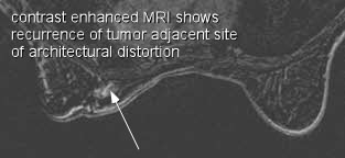
- Evaluating lumpectomy sites in the years following breast cancer treatmentScarring and recurrent cancer can look identical on mammography and ultrasound. If there is a change in a lumpectomy scar by either mammography or on a physical exam, MRI can help determine whether the change is normal maturation of the scar or a recurrence of the cancer.
- Following chemotherapy treatment in patients getting Neoadjuvant ChemotherapyIn some cases, breast cancer will be treated with chemotherapy before it has been removed by surgery. This is called neoadjuvant chemotherapy. In these cases, MRI is often used to monitor how well the chemotherapy is working and to reevaluate the amount of tumor still present before the surgery is performed.
- Evaluating breast implantsMRI is the best test for determining whether silicone implants have ruptured.